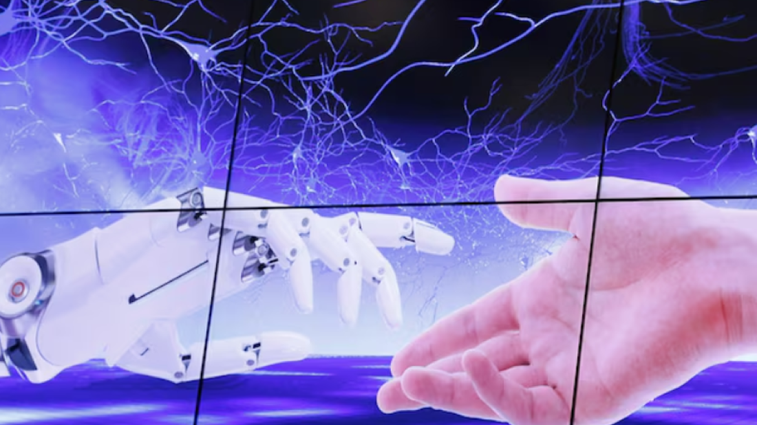
British scientists have built more than 3,800 digital replicas of human hearts using AI and clinical data, aiming to revolutionize diagnosis, correct medical bias, and improve patient care.
In a major leap for personalized medicine, researchers from King’s College London, Imperial College London, and The Alan Turing Institute have developed over 3,800 artificial intelligence-powered digital twins of human hearts.
These hyper-detailed, computer-generated models offer new insights into how heart disease develops and behaves across different ages, sexes, and lifestyles, potentially transforming how patients are diagnosed and treated.
“This is the first time such a large number of patient-specific digital heart models have been created and studied at this scale,” said Steven Niederer, senior author of the study and biomedical engineering chair at Imperial College.
The digital hearts were built by combining AI, medical imaging, and advanced mathematical simulations. A key breakthrough involved training AI to automatically identify and segment heart structures from black-and-white clinical scans. The segmented data was then used to build 3D meshes of individual hearts, which could simulate real-life heart behavior under stress or treatment.
Although running these simulations is computationally demanding—often requiring hours on supercomputers—researchers are now training AI to approximate the results more quickly and affordably.
Challenging medical bias
One of the study’s key findings upends a long-held belief in cardiology: the idea that men and women show inherently different ECG results due to how their hearts function.
“We showed that ECG differences between men and women come down to size, not function,” said Niederer, stressing the importance of reevaluating diagnostic rules that may inadvertently perpetuate gender bias.
The models also offer a way to include underrepresented groups—especially women—in studies that can better predict how they respond to treatments.
Bringing digital hearts into hospitals
While much of the technology is still in development, the team is already working with hospitals in Nottingham and Sheffield to embed the models into clinical workflows.
Supported by The Alan Turing Institute, they are developing cloud-based software that could allow hospitals to upload patient data, generate a digital heart twin in the cloud, and return the results to clinicians in real time.
In future phases, the team hopes to use implantable sensors to feed continuous data into the digital twins, creating dynamic, real-time heart models for ongoing patient monitoring.
From virtual to physical
To make their work more tangible, the team has even begun 3D-printing hearts based on their models. These replicas are being used for education, patient discussions, and even surgical planning.
“This makes what is otherwise a computational object something you can actually hold,” Niederer noted.
Researchers have already expanded the digital twin approach to other medical areas, such as brain tumor modeling in partnership with Cancer Research UK, with a long-term goal of creating whole-body digital twins.
“We’re doing this for the people,” said Cristobal Rodero Gomez of the National Heart and Lung Institute. “Every model, every line of code, is meant to serve a patient.”


What is your opinion on this topic?
Leave the first comment